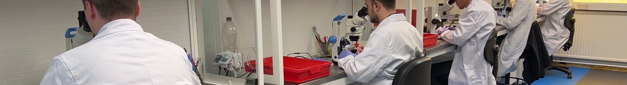
Light Microscopy
Light microscopy is used to make small structures and samples visible by producing a magnified image of their interactions with visible light, such as absorption, reflection, and scattering. This technique helps us understand both the appearance and composition of a sample, and it also allows observation of microscopic processes, for example the diffusion of substances across a cell membrane. By using eyepiece reticles measurements can be determined, counts taken or comparisons made.
There are numerous types of microscopes, with the most common being compound and stereo. A compound microscope has a single optical path whereas a stereo has two optical paths. The types of microscope are more thoroughly explained in the Technology Networks page by Kevin Schädler PhD at this link.
Graticules manufacture five types of products for use with light microscopes: -
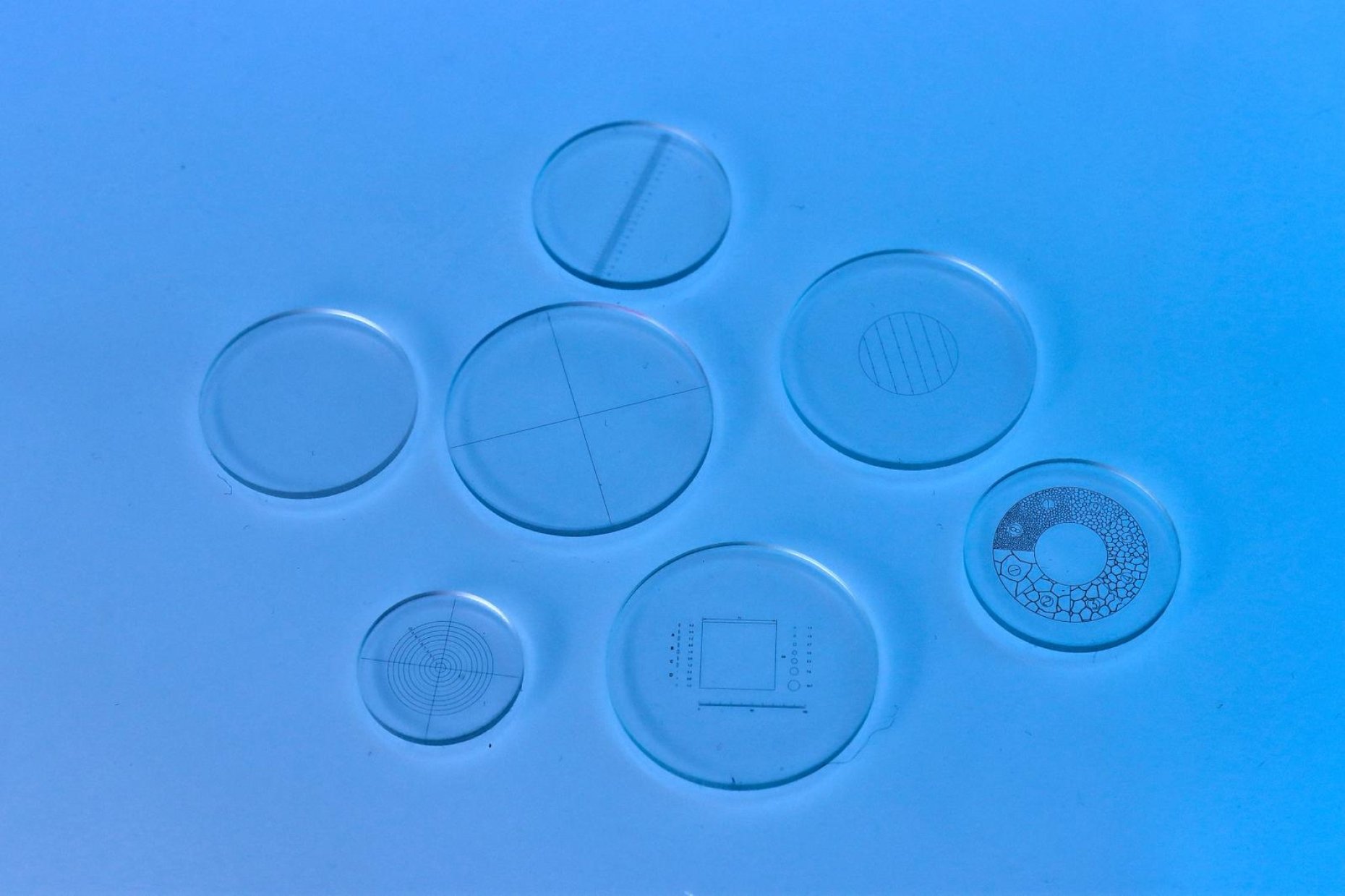
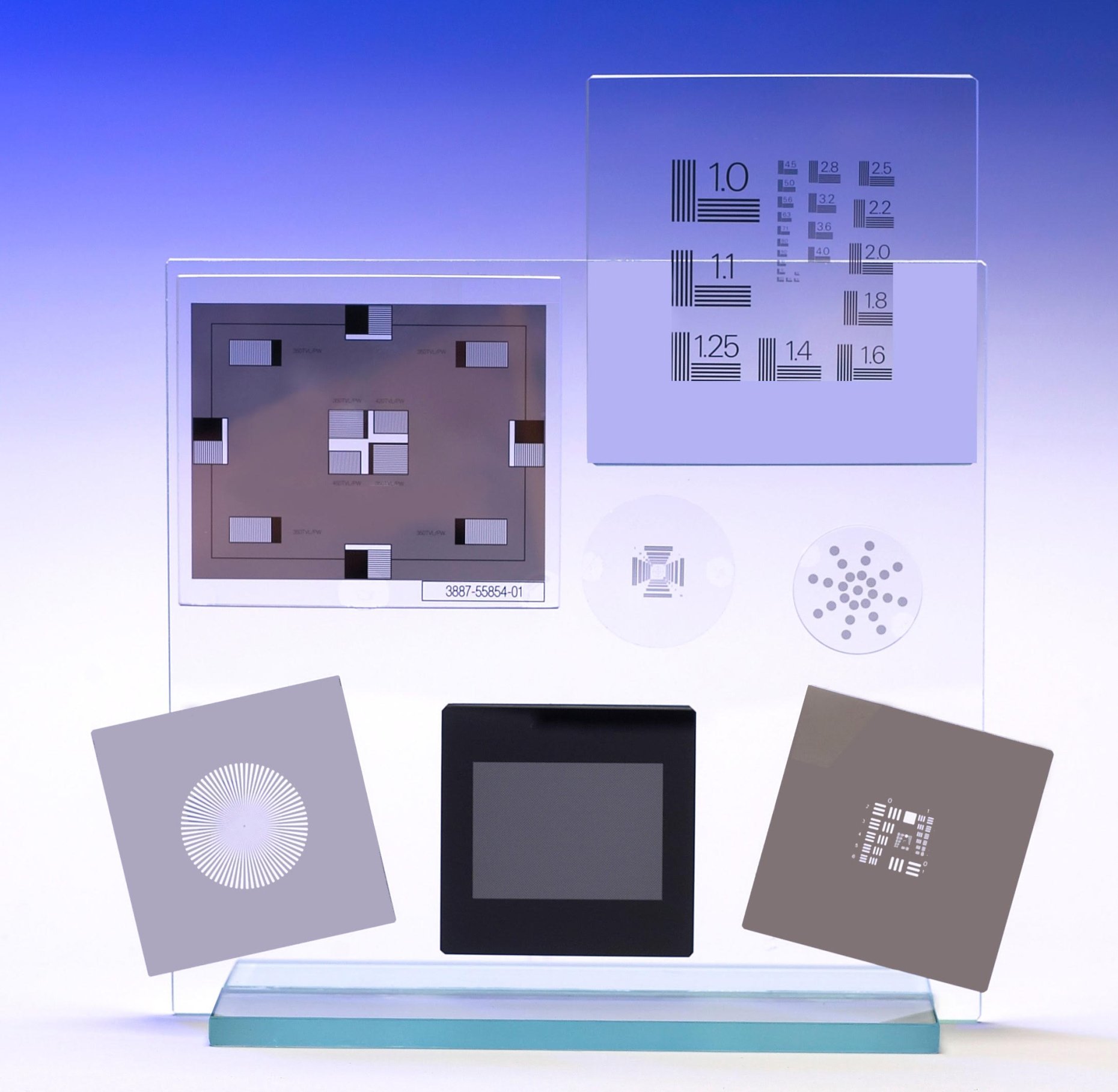
- Eyepiece Reticles (also referred to as eyepiece graticules or ocular micrometers). These are glass disks with a scale, grid or pattern marked on them which fit into the microscope eyepiece/ocular.
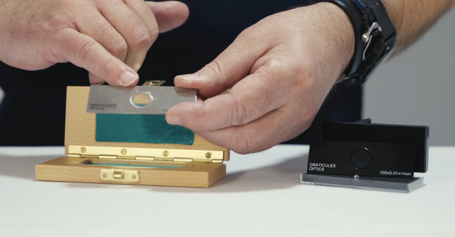
- Stage Micrometers (also referred to as calibration slides or standards). These are used on the stage of the microscope to verify the calibration of the eyepiece reticle or camera imaging system.
- Resolution Slides. These are used to check the quality of the optical system and identify its best resolution.
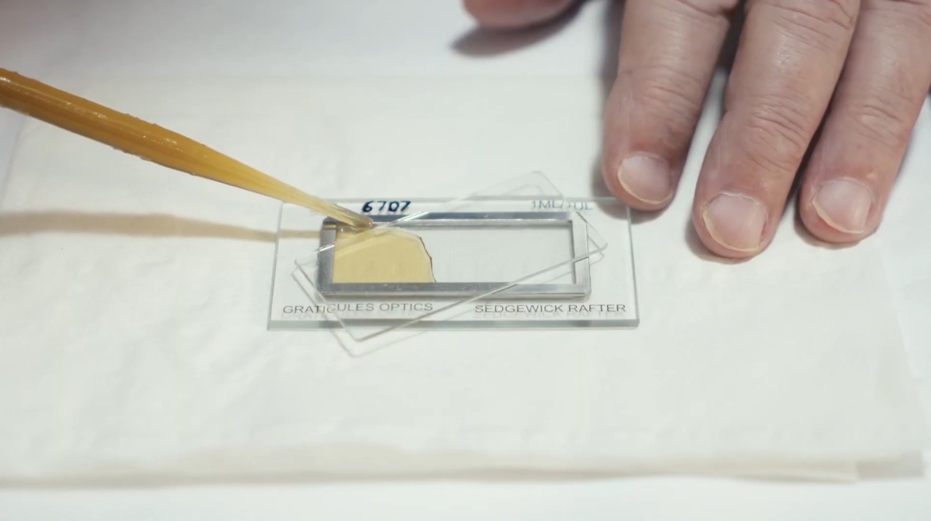
- Counting Chambers. In general, these are stage slides with wells or channels used to analyse liquid specimens. Their uses range from water quality analysis through mold spore detection and sperm examination to blood cell counting. Most have some form of grid pattern either on the base of the slide or on a cover glass in order to provide an accurate count or size measurement.
- Screens. Screen products encompass a wide range of applications, from conductive front glass used in image intensifier systems to viewing screens for profile projectors. Graticules has the capability to apply conductive grid patterns that enable rapid gating of intensifier tubes, a feature commonly required in microscopy, astronomy, and high-speed imaging. In addition, Graticules can supply custom film overlays tailored for use with profile projectors.
Applications of Light Microscopy
Light microscopy has a wide range of applications across many fields, including biology, medicine, forensics, materials science, education, and art conservation. It enables the visualization of specimens ranging from living cells and microorganisms to crystal structures and industrial defects. By employing advanced methods such as fluorescence labelling, three-dimensional imaging, and super-resolution techniques, light microscopy provides detailed insights into both structure and dynamic processes.
Biology and Medicine
In cell biology, light microscopy is used to study living cells, their organelles, cell division, and three-dimensional cell cultures such as organoids. In microbiology, it allows the identification of bacteria, fungi, and parasites based on their morphology and organization. Medical applications include pathology and diagnostics, where biopsy samples, blood smears, and tissue sections are examined to detect disease. In developmental biology, microscopy enables observation of embryonic development and organismal growth, for example in model organisms like zebrafish. It is also used in biochemistry to visualize molecular processes such as protein transport along cytoskeletal structures.
Materials Science and Industry
In materials science, light microscopy is employed to analyse crystal structures, surface features, and defects in metals, ceramics, and semiconductors. In industrial settings, it plays an important role in quality control by inspecting components used in electronics, pharmaceuticals, and manufacturing. It is also valuable in nanotechnology for visualizing nanostructures and studying their behaviour.
Forensics and Environmental Science
Forensic scientists use light microscopy to analyse trace evidence such as hair, fibres, bloodstains, and fingerprints. In environmental science, it is applied to examine soil and water samples, as well as microorganisms within ecosystems.
Art Conservation and Education
In art conservation, microscopy is used to study pigments, fibres, and materials in cultural heritage objects. In education, light microscopy is a fundamental teaching tool, helping students understand concepts ranging from basic cell structure to complex biological processes.
Advanced Microscopy Techniques
Modern light microscopy includes specialized techniques such as fluorescence microscopy, which uses fluorescent markers to track specific molecules and cellular processes in real time. Phase-contrast microscopy enhances the visibility of transparent specimens like living cells. Light-sheet microscopy enables rapid, deep imaging of large samples such as whole embryos or optically cleared tissues, while super-resolution microscopy overcomes the diffraction limit to reveal extremely fine details, including individual proteins.
Product types